Magnetic Resonance Imaging changed what doctors could see without first cutting into the body. Its importance lies not simply in producing pictures, but in turning signals from inside a patient into images that can help guide diagnosis. The supplied account places Sir Peter Mansfield, a London-born physicist working at the University of Nottingham, at the centre of that change. His contribution joined mathematical insight to practical work on nuclear magnetic resonance, helping make a difficult physical phenomenon useful for medical imaging.
MRI is now a familiar part of healthcare, but its origins were not a single sudden invention. The method depended on understanding how nuclei behave in strong magnetic fields, detecting the resulting signals and finding a way to process them into readable pictures. Mansfield’s story is therefore one of scientific translation: from laboratory physics to a technology clinicians could use and patients could experience without an exploratory operation to obtain the first view.
From London education to Nottingham research
Peter Mansfield was born in 1933 and studied at Queen Mary College in London. He joined the University of Nottingham in 1964, entering a research environment in which his work on nuclear magnetic resonance, or NMR, could develop. He was a physicist rather than a clinician: his route into medicine ran through the behaviour of nuclei in magnetic fields and the question of whether the signals they produced could reveal structures inside the human body.
By the early 1970s, Mansfield had made theoretical and practical advances in NMR that helped lay the foundations for MRI. The distinction between those two kinds of work matters. A physical effect may be scientifically interesting, but it does not become a useful diagnostic method simply by being observed. Signals have to be distinguished, measured and interpreted. They must also be organised in a form people can use. Mansfield’s work helped address that problem.
The supplied account identifies a mathematical technique for converting magnetic signals from the body into visual images, with the aid of a computer, as his primary breakthrough. That description captures the essential bridge between magnetic behaviour and a clinical picture. Medicine did not need signals alone; it needed a means of arranging them so that a doctor could interpret what they showed. Mansfield’s contribution helped turn NMR from a specialist physical phenomenon into a basis for imaging.
This was not the work of a lone inventor producing a complete medical machine in one moment. The account describes a series of advances, both theoretical and practical, that helped establish MRI. The achievement depended on making several parts of a process work together: the magnetic conditions, the production and detection of signals, and their mathematical interpretation. The resulting images drew on physics, mathematics and computing, applied to a practical medical need.
How magnetic signals become images
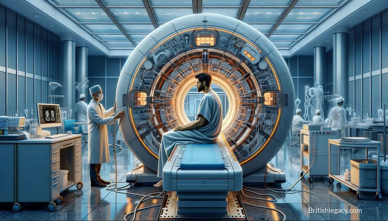
The basic principle begins with nuclear magnetic resonance. In a strong magnetic field, nuclei can absorb and re-emit electromagnetic radiation. MRI uses this behaviour to obtain information from inside the body and build images of its structures. The process can be described in four stages:
- Magnetic field generation: a powerful magnet aligns hydrogen protons in the body.
- Radiofrequency application: a pulse of radio waves disrupts that alignment.
- Signal detection: as the protons return towards their original alignment, they emit signals.
- Image creation: coils detect the signals, and computer processing turns them into pictures of internal structures.
The stages explain why MRI is more than a magnet making a picture. The body’s response produces signals; the machine has to detect them; and computer processing has to turn them into an image that can be interpreted. Mansfield’s work on converting magnetic signals into visual images belongs at that crucial point between the behaviour of the body and the information available to a doctor.
In practical terms, MRI offers a way to examine internal structures without first making an incision. That non-invasive quality changes the diagnostic process: a doctor can obtain an image without exploratory surgery being the means of finding out what is happening inside. The technology’s particular strength, as described in the supplied material, is detailed imaging of soft tissue. That capability has made it useful in several areas of diagnosis rather than merely an impressive demonstration of physics.
A different view for doctors and patients
Before MRI, the search for a diagnosis could involve greater uncertainty and, in some cases, exploratory surgery or exposure to potentially harmful radiation. MRI offered another way to look inside the body. The change was not that every diagnostic problem became straightforward, but that clinicians gained a means of seeing internal structures in detail without the same immediate invasive burden.
The supplied account identifies cancers, neurological disorders and cardiovascular diseases among the conditions for which MRI can assist with early detection. Its soft-tissue imaging is also important in neurological, musculoskeletal and oncological diagnosis. In these fields, the detail in an image can help doctors understand the location and extent of a suspected problem and inform what to do next. The value is practical: better information can support more informed decisions and may help avoid an unnecessary invasive procedure.
Those benefits should not be confused with a claim that a scan alone settles every question. The evidence supplied describes MRI as a diagnostic tool that assists detection and gives detailed images, not as a substitute for clinical judgement. Its significance is that it adds a valuable view of the body to the process of diagnosis. In cases where internal soft tissue matters, that view can make a difference to what doctors can assess.
MRI also developed beyond imaging structure. Functional MRI, or fMRI, uses changes in blood flow to study brain activity. The supplied account notes its contribution to neuroscience and psychology. This extension widened the technology’s reach: magnetic resonance imaging became not only a means of looking for disease or injury, but also a tool for studying activity and function. Its value therefore extends from clinical diagnosis into research on the brain.
Across these uses, the achievement rests on a relationship between technical detail and human need. The scanner gathers signals in a controlled magnetic environment; processing turns them into pictures; clinicians use those pictures to investigate questions about illness and treatment. A development in physics became significant in medicine because the information could be made visible and put to work.
Shared recognition and a wider scientific story
Mansfield was knighted in 1993. In 2003, he shared the Nobel Prize in Physiology or Medicine with Paul Lauterbur, whose related MRI work developed in the United States. The joint award is an important part of the account: MRI’s scientific history includes related work by more than one researcher, and Mansfield’s recognition does not require overlooking Lauterbur’s contribution.
Mansfield’s place in a British account is grounded in the details supplied here: he was born in London, studied at Queen Mary College and carried out the work associated with his career at the University of Nottingham. Those connections provide a clear British setting for his contribution without implying that the development of MRI belonged to one country or one person alone. The shared Nobel recognition places his work within an international scientific achievement while acknowledging the British education and university research behind his part in it.
The most enduring measure of MRI’s reach is its continued use in hospitals around the world. The supplied article states that millions of scans are performed annually. That scale reflects how a specialised line of research became part of routine medical practice. Patients encounter the results not as equations or laboratory signals, but as an examination that can produce detailed images for diagnosis. Clinicians rely on the images to help answer questions that might otherwise remain less clear.
Recognition, then, is not limited to honours. The Nobel Prize and knighthood mark the public standing of Mansfield’s work; continuing hospital use shows its practical reach. MRI links scientific research to decisions made in healthcare, while fMRI extends the same broad technology into research on brain activity. The result is a legacy that spans both the treatment of patients and the study of human function.
Why Magnetic Resonance Imaging deserves a place in British Legacy
Magnetic Resonance Imaging deserves a place in British Legacy because the supplied account shows a specific British contribution to a technology with lasting medical use. Mansfield’s path connects London education at Queen Mary College with research at the University of Nottingham. His work helped convert signals produced through nuclear magnetic resonance into visual images that doctors could interpret. That conversion is the essential achievement: abstract physical behaviour became practical information about the body.
The benefit is not merely technical. MRI allows clinicians to examine internal structures without relying on exploratory surgery to obtain an initial view, and its detailed imaging of soft tissue supports diagnosis across neurological, musculoskeletal and cancer care. The source also describes its use in the assessment of cardiovascular disease and the contribution of fMRI to neuroscience and psychology. These applications show how one body of research can help patients and extend scientific understanding beyond the clinic.
Mansfield shared the 2003 Nobel Prize with Paul Lauterbur, a recognition that rightly situates his British career within a wider international story. Remembering that context makes the British connection more precise, not less significant. MRI’s continuing use in hospitals, and the millions of scans reported annually in the supplied account, demonstrate the enduring reach of the work. Its legacy is a way of seeing inside the body that has become useful in everyday medicine: a union of physics, mathematics and computing directed towards better-informed diagnosis.